 Login / Register


- Regulatory Status
- RUO
- Ave. Rating
- Submit a Review
- Product Citations
- publications

-

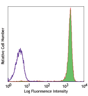
Human peripheral blood mononuclear cells were stimulated with Ultra-LEAF™ Purified anti-human CD3 (clone UCHT1) and Ultra-LEAF™ Purified anti-human CD28 (clone CD28.2) antibodies for 24 hours (filled histogram) or left unstimulated (open histogram) and stained with ATP Red. Data shown were gated on CD3+ lymphocytes. -

Human peripheral blood mononuclear cells were stimulated Ultra-LEAF™ Purified anti-human CD3 (clone UCHT1) and Ultra-LEAF™ Purified anti-human CD28 (clone CD28.2) antibodies for 24 hours. Cells were then treated with Oligomycin A (an ATP synthase inhibitor) (filled histogram) or left untreated (open histogram) and stained with ATP Red. Data shown were gated on CD3+ lymphocytes. -

A549 cells grown on coverslips were treated with 2-Deoxy-D-Glucose (2-DG) (right) for 30 minutes at 37°C or left untreated (control) (left) and subsequently stained with ATP Red for 15 minutes. Cells were then washed with PBS and imaged using a 40X objective.
| Cat # | Size | Price | Quantity Check Availability | Save | ||
|---|---|---|---|---|---|---|
| 421944 | 1 mg | 316€ | ||||
ATP Red is a cell-permeable ATP sensor that localizes to mitochondria and produces fluorescence upon binding to ATP, rapidly responding to fluctuations in mitochondrial ATP levels. It provides a direct and simplified approach to measuring ATP levels in live cells by flow cytometry or live cell imaging. ATP Red allows for the discrimination of rare sub-populations while interrogating important cellular metabolic pathways, such as glycolysis and oxidative phosphorylation. This method is rapid – results can be obtained in less than an hour and yields an accurate measurement of cellular ATP production and its dependency on metabolic pathways of interest.
Product DetailsProduct Details
- Verified Reactivity
- Human, Mouse
- Formulation
- 1 vial of lyophilized ATP Red, 1 mg
- Preparation
-
Bring vial to room temperature. To make a 10 mM stock solution, add 178 µL of DMSO to the vial of 1 mg ATP Red and mix thoroughly until completely dissolved.
Note: Store unused ATP Red stock solution in a tightly sealed vial in the dark at room temperature. Stock solution is stable for at least 12 months. - Storage & Handling
- 2 - 8°C
- Application
-
FC - Quality tested
Live cell imaging - Verified - Recommended Usage
-
Please refer to the detailed protocol in the application notes section.
- Application Notes
-
Components:
1 vial of 1 mg lyophilized ATP Red, Molar Mass: 561.48 g/mol
Storage:
Upon receipt, store lyophilized probe at 2 - 8°C and protected from light. Upon reconstitution, store unused ATP Red stock solution in a tightly sealed vial in the dark at room temperature. Stock solution is stable for at least 12 months.
Imaging Guidelines:
Ex/Em = 590/620 nm
Fluorescence microscope filter set: Cyanine3/TRITC
Flow Guidelines:
Analysis in PE or similar channel (585/42 or 575/25 or 570/40 filter set)
Protocol:- Grow cells in a tissue culture vessel with a coverslip bottom in desired media or prepare cell suspension containing 0.25 - 2.5 x10⁶ cells in 100 - 500 µL of desired media or Cell Staining Buffer (Cat. No. 420201).
- Optional: Fix cells with Fixation Buffer (Cat. No. 420801), if desired.
- Add ATP Red at a final concentration of 5 - 10 µM along with desired antibodies.
- Incubate cells at room temperature or 37°C for 15 minutes (or up to 30 minutes for live cell imaging).
- Optional: Wash cells with PBS or equivalent buffer. If only staining with ATP Red, this washing step can be skipped.
- Proceed directly to analysis.
Antigen Details
- Distribution
-
Mitochondria
- Biology Area
- Adaptive Immunity, Cell Biology, Cell Proliferation and Viability, Immunology, Innate Immunity, Mitochondrial Function
- Molecular Family
- Mitochondrial Markers
- Antigen References
-
- Wang L, et al. 2016. Angew Chem Int Ed Engl. 55:1773-6.
- Gene ID
- NA


Follow Us