 Login / Register


 United States ($) USD
 Austria (€) EUR
 Belgium (€) EUR
 Denmark (DKK) DKK
 Finland (€) EUR
 France (€) EUR
 Germany (€) EUR
 Ireland (€) EUR
 Japan (¥) JPY
 Luxembourg (€) EUR
 Netherlands (€) EUR
 Norway (NOK) NOK
 Sweden (SEK) SEK
 Switzerland (CHF) CHF
 United Kingdom (£) GBP

- Clone
- 16A1 (See other available formats)
- Regulatory Status
- RUO
- Other Names
- Platelet-derived growth factor receptor, alpha polypeptide, PDGFR2, PDGFRα, PDGFRa, PDGF receptor alpha
- Isotype
- Mouse IgG1, κ
- Ave. Rating
- Submit a Review
- Product Citations
- publications
The 16A1 monoclonal antibody recognizes human CD140a also known as the platelet-derived growth factor receptor, alpha polypeptide, PDGFR2, and PDGFRα. CD140a is a cell surface tyrosine kinase receptor for members of the platelet-derived growth factor family. The identity of the growth factor bound to the receptor determines whether the functional receptor is a homodimer or heterodimer composed of both PDGFR-α and -β. CD140a contains three immunoglobulin-like domains and a tyrosine kinase domain with a predicted molecular weight of approximately 123 kD. CD140a is widely expressed on a variety of mesenchymal-derived cells and has been implicated in the development of some tumors including basal cell carcinoma and gastric stromal cell tumors. Binding of A-chain containing PDGF molecules as well as protease-activated PDGF-C molecules can stimulate cell proliferation. CD140a has been shown to interact with a number of proteins including CRK, Grb2, Grb14, SHP2, and others as integrin β3, caveolin-1, and nexin sorting molecules. The PDGFRα is heavily phosphorylated on numerous tyrosine residues through both autophosphorylation and ligand-dependent processes. The 16A1 antibody has been shown to be useful for flow cytometric detection of CD140a.
Product DetailsProduct Details
- Verified Reactivity
- Human
- Antibody Type
- Monoclonal
- Host Species
- Mouse
- Immunogen
- NIH 3T3 cells transfected with human PDGFRalpha
- Formulation
- Phosphate-buffered solution, pH 7.2, containing 0.09% sodium azide
- Preparation
- The antibody was purified by affinity chromatography and conjugated with Spark PLUS UV395™ under optimal conditions.
- Concentration
- 0.2 mg/mL
- Storage & Handling
- The antibody solution should be stored undiluted between 2°C and 8°C, and protected from prolonged exposure to light. Do not freeze.
- Application
-
FC
- Recommended Usage
-
Flexi-Fluors™ are provided at a standard 0.2 mg/mL concentration. We recommend titrating this reagent to determine the optimal concentration for each application. For many flow cytometry applications, conjugated antibodies perform well at concentrations ranging from 0.03 to 1.0 µg per million cells in 100 µL. We recommend testing a range of concentrations starting from 10 µg/mL.
For example, make five 1:1 serial dilutions of the 0.2 mg/mL antibody. Add 5 µL of each dilution (including the undiluted antibody) to 100 µL of cells (at 107 cells/mL) to test six concentrations -- 1.0, 0.5, 0.25, 0.125, 0.06, and 0.03 µg per million cells in 100 µL volume. Compare staining patterns or create a titration curve using the MFI or staining index to determine the optimal concentration.
* Spark PLUS UV395™ has a maximum excitation of 355 nm and a maximum emission of 385 nm. - Excitation Laser
-
Ultraviolet Laser (355 nm)
- Additional Product Notes
-
For more information about Flexi-Fluors™, visit our Flexi-Fluor™ page and review FAQs associated with this product line.
-
Application References
(PubMed link indicates BioLegend citation)
Antigen Details
- Structure
- Cell surface tyrosine kinase receptor for members of the platelet-derived growth factor family.
- Distribution
-
Widely expressed on a variety of mesenchymal-derived cells.
- Function
- Stimulation of cell proliferation; mitogenic activity for cells of mesenchymal origin. Knock-out studies have implicated an essential role for CD140a in kidney development. Has been implicated in basal cell carcinoma and gastric stromal cell tumors.
- Interaction
- Interacts with Crk, as well as a variety of adaptor molecules and signaling intermediates (Grb2, Grb14, SHP2, others). Has also been shown to associate with integrin β3, caveolin-1, and nexin sorting molecules
- Ligand/Receptor
- Binds to A-chain containing PDGF molecules and protease-activated PDGF-C molecules
- Modification
- Multiple tyrosine phosphorylation sites (Y720, Y731, Y742, Y754, Y762, Y767, Y768, Y988, Y993, Y1018)
- Cell Type
- Embryonic Stem Cells, Mesenchymal cells, Mesenchymal Stem Cells, Neural Stem Cells
- Biology Area
- Angiogenesis, Cell Biology, Immunology, Neuroscience, Neuroscience Cell Markers, Stem Cells
- Molecular Family
- CD Molecules, Cytokine/Chemokine Receptors
- Antigen References
-
1. Gronwald RG, et al. 1988. Proc. Natl. Acad. Sci. USA 85:3435.
2. Gilbertson DG, et al. 2001. J. Biol. Chem. 276:27406.
3. Seifert RA, et al. 1989. J. Biol. Chem. 264:8771.
4. Rupp E, et al. 1994. Eur. J. Biochem. 225:29. - Gene ID
- 5156 View all products for this Gene ID
- UniProt
- View information about CD140a on UniProt.org


Related FAQs
- What are Flexi-Fluors?
-
Flexi-Fluors are rapidly made-to-order conjugated antibodies. The technology, manufacturing processes, and specifications used to create Flexi-Fluors are the same as our regular catalog products. However, the optimal concentration and performance of each Flexi-Fluor must be determined by the customer.
- How quickly will I receive my order?
-
We aim to ship Flexi-Fluors within 2-3 weeks of receipt of your order. However, depending on your location, shipping times may vary.
- How are Flexi-Fluors different from regular catalog products?
-
Flexi-Fluors are made on demand, specifically for you. Flexi-Fluors are manufactured using the same high-quality standards, and specifications as other catalog products. For faster delivery, Flexi-Fluors are not tested by flow cytometry to determine optimal concentrations or evaluate performance. This testing needs to be performed by the customer.
- How do I determine the optimal concentration for using my Flexi-Fluor? How should I titrate my antibody?
-
Flexi-Fluors are provided at a standard 0.2 mg/mL concentration. We recommend that you titrate your antibody to determine the optimal concentration to use for your application. For many flow cytometry applications, conjugated antibodies perform well at concentrations ranging from 0.03 to 1.0 µg per million cells in 100 µL volume. We recommend that you test a range of concentrations starting from 10 µg/mL.
For example, make five 1:1 serial dilutions of your 0.2 mg/mL antibody. Add 5 µL of each dilution (including the undiluted antibody) to 100 µL of cells (at 107 cells/ml) to test six concentrations - 1.0, 0.5, 0.25, 0.125, 0.06, and 0.03 µg per million cells in 100 µL volume. Compare staining patterns or create a titration curve using the MFI or staining index to determine the optimal concentration.
- I can’t find the antibody-dye combination that I need. When will it be available?
-
We continuously update our catalog, introducing scores of new products every month. Please get in touch with our Technical Service team for an update on new products or recommendations for suitable alternatives to complete your panel. Or contact Custom Solutions to inquire about our affordable custom conjugation services.
- I need help to validate the performance of my Flexi-Fluor. Who should I contact?
-
Please get in touch with Technical Service for assistance.
- Can I order more than 50 μg of a Flexi-Fluor?
-
Yes, you can order multiple vials of the same Flexi-Fluor products. We cannot guarantee, however, that these vials will be bottled from the same lot. For bulk single-lot orders, contact our Custom Solutions team.
- What is the expiration date of my Flexi-Fluor?
-
Expiration dates can be found on the vial label or by using our CoA lookup tool.
- What if my requested antibody-dye combination doesn't perform as expected?
-
Flexi-Fluor™ products have been carefully selected by our scientists for their excellent conjugation properties and broad compatibility. In the rare case that an antibody-dye combination does not perform as expected due to the unique characteristics of a specific pairing, please contact our Technical Service team. We will work with you to identify alternative options to meet your panel requirements.
Other Formats
View All CD140a Reagents Request Custom Conjugation| Description | Clone | Applications |
|---|---|---|
| PE/Cyanine7 anti-human CD140a (PDGFRα) | 16A1 | FC |
| TotalSeq™-A0128 anti-human CD140a (PDGFRα) | 16A1 | PG |
| APC anti-human CD140a (PDGFRα) | 16A1 | FC |
| TotalSeq™-C0128 anti-human CD140a (PDGFRα) | 16A1 | PG |
| TotalSeq™-B0128 anti-human CD140a (PDGFRα) | 16A1 | PG |
| Purified anti-human CD140a (PDGFRα) | 16A1 | FC,ICC |
| Biotin anti-human CD140a (PDGFRα) | 16A1 | FC |
| PE anti-human CD140a (PDGFRα) | 16A1 | FC |
| TotalSeq™-D0128 anti-human CD140a (PDGFRα) | 16A1 | PG |
| KIRAVIA Blue 520™ anti-human CD140a (PDGFRα) Antibody | 16A1 | FC |
| Spark PLUS UV395™ anti-human CD140a (PDGFRα) (Flexi-Fluor™) | 16A1 | FC |
Compare Data Across All Formats
This data display is provided for general comparisons between formats.
Your actual data may vary due to variations in samples, target cells, instruments and their settings, staining conditions, and other factors.
If you need assistance with selecting the best format contact our expert technical support team.
-
PE/Cyanine7 anti-human CD140a (PDGFRα)
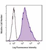
Human PDGFRα transfected NIH/3T3 cells were stained wi... -
TotalSeq™-A0128 anti-human CD140a (PDGFRα)
-
APC anti-human CD140a (PDGFRα)
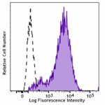
Human PDGFRα transfected NIH/3T3 cells were stained with CD1... -
TotalSeq™-C0128 anti-human CD140a (PDGFRα)
-
TotalSeq™-B0128 anti-human CD140a (PDGFRα)
-
Purified anti-human CD140a (PDGFRα)
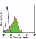
Human PDGFRA transfected cells stained with purified 16A1, f... -
Biotin anti-human CD140a (PDGFRα)
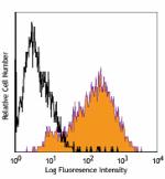
Human PDGFRA transfected cells stained with biotinylated 16A... -
PE anti-human CD140a (PDGFRα)
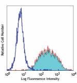
Human PDGFRA transfected cells stained with16A1 PE -
TotalSeq™-D0128 anti-human CD140a (PDGFRα)
-
KIRAVIA Blue 520™ anti-human CD140a (PDGFRα) Antibody

Human PDGFRα transfected NIH/3T3 cells were stained with ant... -
Spark PLUS UV395™ anti-human CD140a (PDGFRα) (Flexi-Fluor™)


Follow Us