 Login / Register


 United States ($) USD
 Austria (€) EUR
 Belgium (€) EUR
 Denmark (DKK) DKK
 Finland (€) EUR
 France (€) EUR
 Germany (€) EUR
 Ireland (€) EUR
 Japan (¥) JPY
 Luxembourg (€) EUR
 Netherlands (€) EUR
 Norway (NOK) NOK
 Sweden (SEK) SEK
 Switzerland (CHF) CHF
 United Kingdom (£) GBP

- Clone
- D6E10 (See other available formats)
- Regulatory Status
- RUO
- Other Names
- Apolipoprotein E, apolipoprotein E3, AD2, LDLCQ5, LPG, ApoE
- Previously
-
Signet Catalog# 9740-02
Signet Catalog# 9740-05
Signet Catalog# 9740-10
Covance Catalog# SIG-39740
- Isotype
- Mouse IgG1, κ
- Ave. Rating
- Submit a Review
- Product Citations
- publications

-

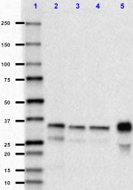
Western blot of purified anti-Apo E antibody (clone D6E10). Lane 1: Molecular weight marker; Lane 2: 40 ng of Apo E2 recombinant protein; Lane 3: 40 ng of Apo E3 recombinant protein; Lane 4: 40 ng of Apo E4 recombinant protein; Lane 5: 40 µg of human plasma. The blot was incubated with 1 µg/mL of the primary antibody overnight at 4°C, followed by incubation with HRP labeled goat anti-mouse IgG (Cat. No. 405306). Enhanced chemiluminescence was used as the detection system. -
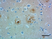
IHC staining of purified anti-Apo E antibody (clone D6E10) on formalin-fixed paraffin-embedded Alzheimer’s disease brain tissue. Following antigen retrieval using Sodium Citrate H.I.E.R., the tissue was incubated with 5 µg/mL of the primary antibody overnight at 4°C. BioLegend's Ultra Streptavidin (USA) HRP Detection Kit (Multi-Species, DAB, Cat. No. 929901) was used for detection followed by hematoxylin counterstaining, according to the protocol provided. The image was captured with a 40X objective. Scale bar: 50 µm -
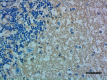
IHC staining of purified anti-Apo E antibody (clone D6E10) on formalin-fixed paraffin-embedded human cerebellum tissue. Following antigen retrieval using Sodium Citrate H.I.E.R., the tissue was incubated with 10 µg/mL of the primary antibody overnight at 4°C. BioLegend's Ultra Streptavidin (USA) HRP Detection Kit (Multi-Species, DAB, Cat. No. 929901) was used for detection followed by hematoxylin counterstaining, according to the protocol provided. The image was captured with a 40X objective. Scale bar: 50 µm -
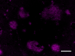
IHC staining of purified anti-Apo E antibody (clone D6E10) antibody on formalin-fixed paraffin-embedded Alzheimer’s disease brain tissue. Following antigen retrieval using Sodium Citrate H.I.E.R., the tissue was incubated with 10 µg/mL of the primary antibody overnight at 4°C, followed by incubation with Alexa Fluor® 647 goat anti-mouse IgG (Cat No. 405322) for one hour at room temperature. The image was captured with a 40X objective. Scale bar: 50 µm -

HepG2 cells (negative cell line, panel A) and HeLa cells (positive cell line, panel B) were fixed with 4% PFA Fixation Buffer (Cat. No. 420801) for 10 minutes, permeabilized with 0.5% Triton-X100 for 10 minutes, and blocked with 5% FBS for 1 hour at room temperature. Cells were then stained with Purified anti-Apo E (clone D6E10) and Flash PhalloidinTM Green 488 (green, Cat. No. 424201), followed by incubation with Alexa Fluor® 594 Goat anti-mouse IgG (red, Cat. No. 405326) for 1 hour at room temperature. Nuclei were counterstained with DAPI (Cat. No. 422801). The images were captured on a Revvity Operetta CLS™ High Content Analysis System with a 63X objective and merged. Scale bar: 50 μm
| Cat # | Size | Price | Quantity Check Availability | Save | ||
|---|---|---|---|---|---|---|
| 803303 | 1 mL | 1399€ | ||||
| 803301 | 200 µL | 449€ | ||||
Apo-E is a class of apolipoprotein primarily synthesized in the liver, but is also found in other tissues such as brain, kidneys, and spleen. Apo-E is a protein component of serum chylomicron and lipoprotein particles, playing essential roles in cholesterol transportation, homeostasis, and metabolism. The expression of Apo-E is induced by cholesterol-rich diets and hyperlipoproteinemia. In the central nervous system, Apo-E is produced by astrocytes and microglia. The Apo-E-containing lipoprotein particles play important functions in brain injury repair, neurogenesis, and synaptic plasticity. Apo-E has been found to be involved in the regulation of amyloid-β aggregation and clearance. Genetic polymorphisms of Apo-E are associated with neuron degenerative disorders including Alzheimer's disease.
Product DetailsProduct Details
- Verified Reactivity
- Human
- Antibody Type
- Monoclonal
- Host Species
- Mouse
- Immunogen
- This antibody was raised against a peptide sequence corresponding to aa 141-160 of human Apo-E.
- Formulation
- Phosphate-buffered solution (no preservatives or carrier proteins).
- Preparation
- The antibody was purified by affinity chromatography.
- Concentration
- 1 mg/ml
- Storage & Handling
- This antibody should be handled aseptically as it is free of preservatives such as Sodium Azide. Store this antibody undiluted between 2°C and 8°C. Please note the storage condition for this antibody has been changed from -20°C to between 2°C and 8°C. You can also check the vial label or CoA to find the proper storage conditions.
- Application
-
WB - Quality tested
IHC-P, ICC - Verified - Recommended Usage
-
Each lot of this antibody is quality control tested by Western blotting. For Western blotting, the suggested use of this reagent is 0.5 - 1.0 µg/mL. For immunohistochemistry on formalin-fixed paraffin-embedded tissue sections, a concentration range of 5 - 10 µg/mL is suggested. For immunocytochemistry, a concentration range of 1.0 - 10 is suggested. It is recommended that the reagent be titrated for optimal performance for each application.
- Additional Product Notes
-
For use in immunocytochemistry (ICC), it is recommended to fix/permeabilize with any of the following:
-Fixation buffer (Cat. No. 420801) with 0.5 % Triton X-100
-Fixation buffer (Cat. No. 420801) with 100% ice-cold methanol
-100% ice-cold methanol only
Fixation and permeabilization with 100% ice-cold methanol results in dimmer signal compared to the other methods.
This product is not recommended for use in intracellular flow cytometry (ICFC) -
Application References
(PubMed link indicates BioLegend citation) -
- Munoz DG and Feldman H. 2000. CMAJ. 162(1):65-72.
- Hagberg JM, et al. 2000. Physiol Genomics. 4(2):101-108.
- Saunders AM. 2000. J Neuropathol Exp Neurol. 59(9):751-8.
- Golabek AA, et al. 2000. Biophys J. 79(2):1008-15.
- Zunarelli E, et al. 2000. Breast Cancer Res Treat. 63(3):193-8.
- Zunarelli E, et al. 2000. Clin Neuropathol. 19(1):1-6.
- Tomiyama T, et al. 1999. Cell Mol Life Sci. 6(3-4):268-79.
- Golabek AA., et al. 1996. J Biol Chem. 271(18):10602-6.
- Product Citations
-
- RRID
-
AB_2564661 (BioLegend Cat. No. 803303)
AB_2564662 (BioLegend Cat. No. 803301)
Antigen Details
- Biology Area
- Cell Biology, Neurodegeneration, Neuroscience, Protein Trafficking and Clearance
- Gene ID
- 348 View all products for this Gene ID
- UniProt
- View information about Apo E on UniProt.org


Related FAQs
Other Formats
View All Apo E Reagents Request Custom Conjugation| Description | Clone | Applications |
|---|---|---|
| Purified anti-Apo E | D6E10 | WB,IHC-P,ICC |
| Biotin anti-Apo E | D6E10 | WB |
| Alexa Fluor® 647 anti-ApoE Antibody | D6E10 | IHC-P,ICC |
Customers Also Purchased
Compare Data Across All Formats
This data display is provided for general comparisons between formats.
Your actual data may vary due to variations in samples, target cells, instruments and their settings, staining conditions, and other factors.
If you need assistance with selecting the best format contact our expert technical support team.
-
Purified anti-Apo E
Western blot of purified anti-Apo E antibody (clone D6E10). ... 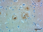
IHC staining of purified anti-Apo E antibody (clone D6E10) o... 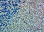
IHC staining of purified anti-Apo E antibody (clone D6E10) o... 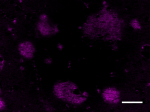
IHC staining of purified anti-Apo E antibody (clone D6E10) a... 
HepG2 cells (negative cell line, panel A) and HeLa cells (po... -
Biotin anti-Apo E
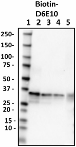
Western blot of Biotin anti-Apo E antibody (clone D6E10). La... -
Alexa Fluor® 647 anti-ApoE Antibody

IHC staining with Alexa Fluor® 647 anti-ApoE (clone D6E10) o... 
HeLa cells (negative cell line, panel A) and HepG2 cells (po...


Follow Us