 Login / Register


- Regulatory Status
- RUO
- Other Names
- FQ6, LysoFQ-GBA, GBA substrate, GBA fluorescent substrate, GCase probe
- Ave. Rating
- Submit a Review
- Product Citations
- publications

-

-

GCase/GBA1 activity detection on the indicated cell lines using LysoFQ-GBA (Lysosomal GCase Probe Green) live cell imaging (green) and Hoechst nuclear staining (blue). -

Time-course analysis of GCase activity in A549 cells was conducted by incubating the cells with LysoFQ-GBA (Lysosomal GCase Probe Green) for 2 hours. Following incubation, cells were washed and resuspended in phenol-free media. Live-cell imaging was performed over an 8-hour period, with representative images captured at 0, 4, and 8 hours using a Revvity Operetta CLS™ High Content Analysis System equipped with a 63X objective. Hoechst staining (blue) was used for nuclear visualization. -

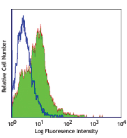
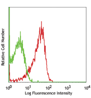
A-549 cells untreated (black line histogram) or treated with the LysoFQ-GBA (Lysosomal GCase Probe Green) (purple filled histogram) for 2 hours. The cells were fixed using Fixation Buffer (Cat. No. 420801) and analyzed by flow cytometry using a FITC channel.
| Cat # | Size | Price | Quantity Check Availability | Save | ||
|---|---|---|---|---|---|---|
| 421948 | 500 µg | 200€ | ||||
Important Note: This product is available for strictly non-commercial use to researchers employed by or affiliated with academic institutions, non-profit organizations, and for-profit companies. For any requests for commercial use licenses, please have representatives from the respective academic institutions, non-profit organizations, and for-profit companies contact our Business Development group. Please be advised that in an effort to prevent delays in order fulfillment and maintain reagent accessibility for all researchers, orders of this reagent may be limited to a total quantity of 10 vials per month. For questions or exemption requests, please contact your local BioLegend representative.
The enzyme glucocerebrosidase/GCase (encoded by the GBA1 gene) is essential for the hydrolysis of glucocerebroside, a lipid substrate within lysosomes. This enzymatic function is critical for maintaining cellular lipid metabolism and lysosomal integrity. Mutations in the GBA1 gene can result in diminished or defective glucocerebrosidase activity, leading to the intracellular accumulation of glucocerebroside. This accumulation is the primary etiological factor in Gaucher disease, a lysosomal storage disorder manifesting with hepatosplenomegaly, skeletal abnormalities, and neurological complications. Furthermore, GBA1 mutations are recognized as significant genetic risk factors for Parkinson's disease, as they compromise lysosomal function and disrupt cellular homeostasis, thereby contributing to neurodegenerative processes.
This LysoFQ-GBA (Lysosomal GCase Probe Green) probe, a cell-permeable reporter for GCase activity, fluoresces upon glucocerebrosidase activation, facilitating the detection and monitoring of GBA1 activity. This is pivotal for the diagnosis and management of related diseases, enabling early diagnosis, elucidation of disease progression, and the development of targeted therapies. Thus, comprehending the function and activity of GBA1 is imperative for advancing therapeutic strategies and improving disease management for these conditions. Additionally, a correlation between GCase activity in iPSC-derived neurons and monocytes suggests that patient blood samples may serve as a surrogate for GCase activity in patient neurons.
Product Details
- Verified Reactivity
- Human
- Formulation
- Lyophilized
- Preparation
- See application notes.
- Storage & Handling
- -70°C
- Application
-
ICC, Live cell imaging, FC - Verified
- Recommended Usage
-
FC, ICC & Live cell staining: See Application Notes
- Application Notes
-
Component:
500 µg of LysoFQ-GBA (Lysosomal GCase Probe Green)
General Considerations:- Optimal concentration and loading time will vary depending on cell type, application and experimental conditions. Recommended working concentrations range between 2.5 -20 μM LysoFQ-GBA (Lysosomal GCase Probe Green).
- LysoFQ-GBA (Lysosomal GCase Probe Green) is compatible with kinetic imaging and analysis for up to 8 hours in cell culture and under appropriate conditions.
- AT3375 and isofagomine are potent Glucocerebrosidase inhibitors that can be used for control samples. An effective AT3375 and isofagomine concentration is typically 10 μM for overnight incubation at 37°C prior to LysoFQ-GBA (Lysosomal GCase Probe Green) addition for most cell types. We recommend users to determine optimal conditions based on their assay.
Reconstitution and Preparation of Stock Solution:
- Thaw and bring LysoFQ-GBA (Lysosomal GCase Probe Green) to room temperature in dark for at least 10 mins
- Add 77.3 µL of DMSO to make a stock solution of 5 mM. Vortex (maximum speed) for 60s and briefly centrifuge.
- Aliquot the stock immediately into several one-time-use tubes of desired volumes. Avoid freeze thaw cycles with this material and always keep in the dark.
- We do not recommend re-freezing any unused stock solution after a single aliquot has been thawed for experimental use.
Preparation of Working Solution:
- Allow the aliquot of LysoFQ-GBA (Lysosomal GCase Probe Green) stock solution to thaw and reach room temperature protected from light.
- Determine the desired working concentration and volume of LysoFQ-GBA (Lysosomal GCase Probe Green) for your assay.
- Prepare the working solution (between 2.5 -20 μM) by diluting the LysoFQ-GBA (Lysosomal GCase Probe Green) stock solution with the desired assay buffer (such as phenol red-free culture media or PBS).
Microscopy assay (live cell imaging and ICC)
This protocol is designed for adherent cell lines. It is recommended to use surface coating, such as poly-L-Lysine, to promote cell adhesion and avoid washing off cells during wash steps. If working will cells in suspension, optimal assay conditions may need to be determined.- Gently remove media from cells which were seeded onto the desired live cell imaging plates.
- Gently add the working solution of LysoFQ-GBA (Lysosomal GCase Probe Green). Incubate at 37°C for 2 hours and protected from light.
- Wash wells with an excess of phenol red-free medium or buffer of choice. Repeat wash 2 times for a total of 3 washes. Be careful not to disrupt cell adhesion.
- Add the desired amount of phenol red-free medium supplemented with 2-10% FBS.
- Perform live cell imaging using the FITC channel for LysoFQ-GBA (Lysosomal GCase Probe Green) detection.
Flow cytometry assay
- Prepare a 1X working solution of LysoFQ-GBA (Lysosomal GCase Probe Green) by diluting the stock solution to 5 µM in the desired phenol red-free media or buffer containing 5% FBS.|
Note: The working concentration of 5 µM for 2 hours was optimized for human PBMCs and CD14+ monocytes. It may be necessary to determine the optimal concentrations for the desired assay conditions and cell types. - For adherent cells, gently remove culture media and replace with 1X working solution of LysoFQ-GBA (Lysosomal GCase Probe Green) and incubate for at least 2 hours at 37°C and 5% CO2.
Note: multiple time points may be desired depending on your experimental design. - Then, gently remove the solution from cells and replace with phenol red-free media containing 5% FBS.
- Harvest the cells following desired detachment reagent (such as trypsin) and resuspend in the desired flow cytometry buffer/solution.
Note: Cells can be fixed with PFA 2% or Fixation Buffer (Cat. No. 420801) for 10 min. - Analyze the cells using a flow cytometer using a FITC or equivalent channel for detection of LysoFQ-GBA (Lysosomal GCase Probe Green).
Detection/Imaging Guidelines
- Ex/Em: 503/509
- Fluorescence microscope filter set: FITC
- Flow Cytometer Channel: FITC - Additional Product Notes
-
This product is intended for single use after reconstitution and performance is affected by frozen-thaw cycles. If more than single use is needed, we recommend store reconstituted product in single use aliquots at -70°C (Please see application notes for details).
- Application References
-
- Deen MC, et al. 2022. Proc Natl Acad Sci U S A.119:29.
Antigen Details
- Structure
-
Chemical Formula: C63H84BF2N15O
Molecular weight: 1292.26 g/mol - Antigen References
-
- Deen MC, et al. 2022. Proc Natl Acad Sci U S A.119:29.
- Gehrlein A, et al. 2023. Nature Communications. 14:2057
- Zhu S, et al. 2023. Angew Chem Int Ed Engl. 62,e202309306.
- Williams D, et al. 2024. Proc Natl Acad Sci U S A.
- Koros C, et al. 2024. Genes (Basel). 16; 15:1605.
- Gene ID
- NA


Follow Us